Exhibition
Neuroscience has an artistic dimension.
This exhibition displays a selection of scientific images produced by researchers of the Human Brain Project (HBP) in recent years.
The images portray the same subject - the human brain. Some are extremely detailed photographs, others are digital renderings or 3D simulations. None of the images have been originally created with an artistic intent, but have been repurposed as such, to be displayed as if they were part of an art gallery. A scientific description of the image has been included, both in English and French.
The scientists who created these pictures belong to the many academic institutions which were part of the HBP. The brain research displayed here now continues within the EBRAINS research infrastructure.
We would like to invite you to explore the artistic essence of neuroscience and wish you an enjoyable visit into the brain.
ALPHABET
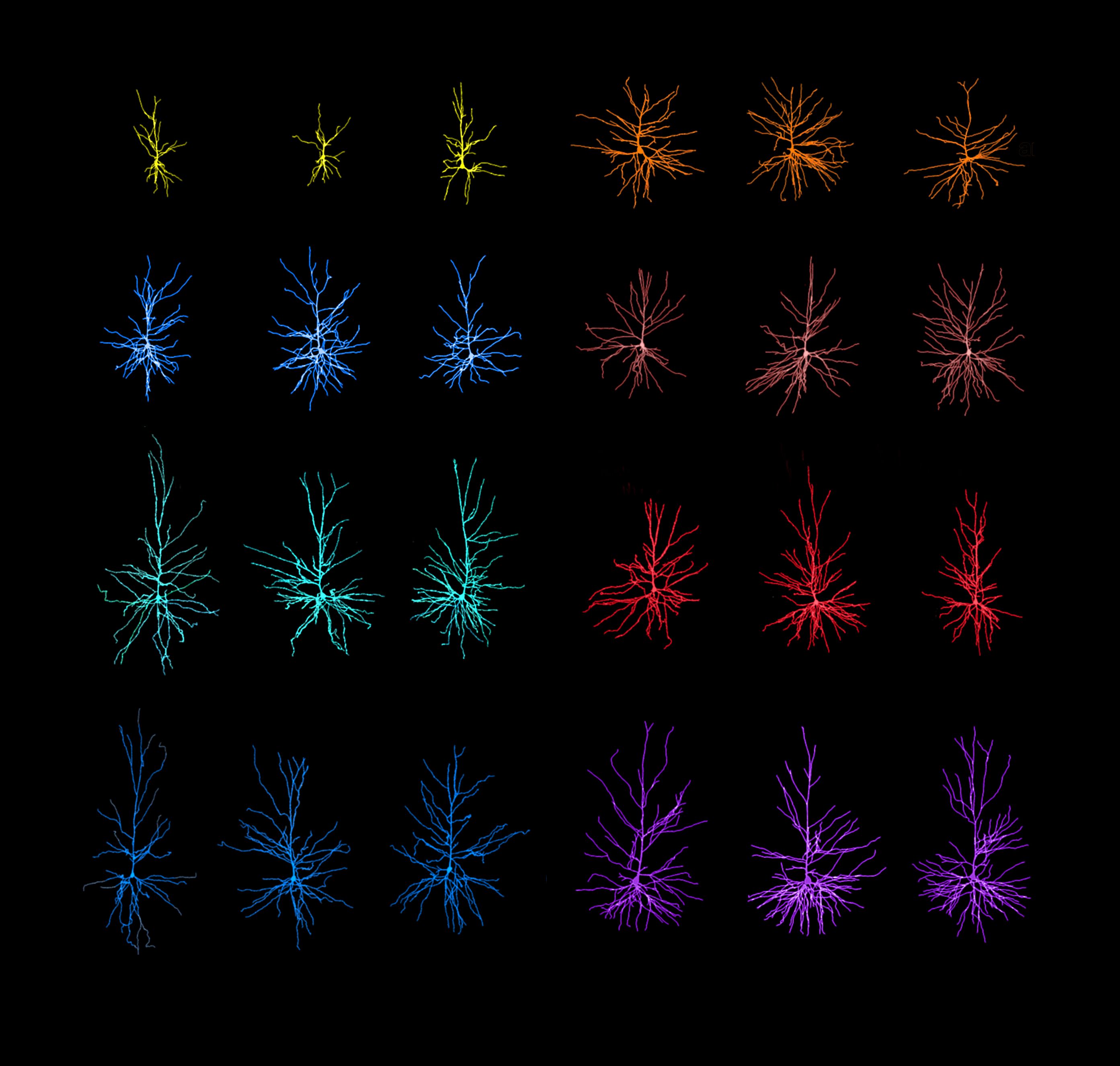
Drawings of layer III pyramidal neurons from different regions of the Brodmann's area (BA) 17 (yellow), BA4 (light blue), BA24 (green), BA10 (dark blue), BA22 (orange), BA21 (salmon), BA20 (red) and BA9 (purple) of the human cerebral cortex. Note the large variations in the pyramidal cell structure depending on the cortical region.
EYE
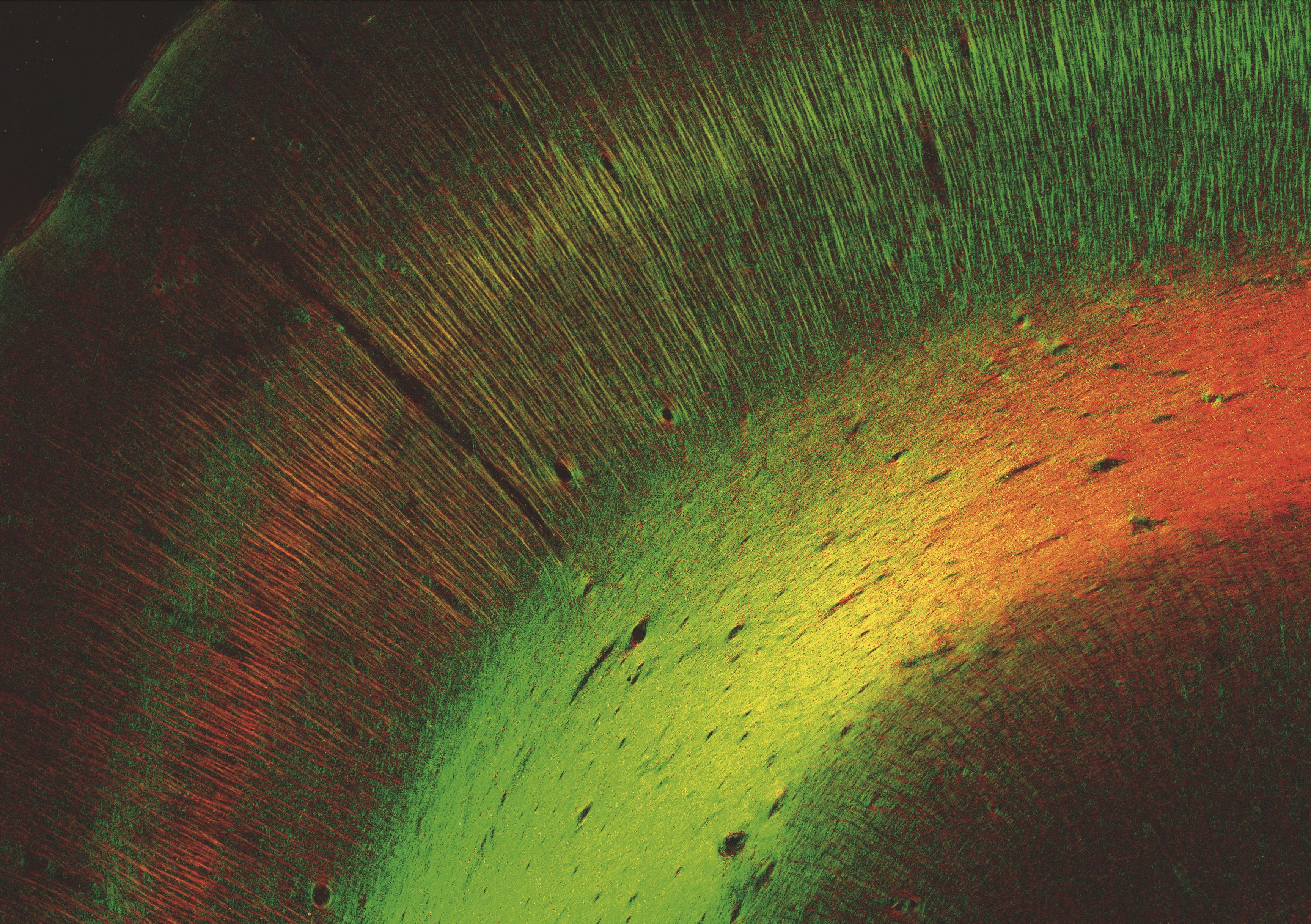
Fiber orientation map of the human visual cortex revealed by 3D Polarized Light Imaging. Determined in-plane orientations are coded in RGB color space (red: x-direction, green: y-direction).
MEMORIES
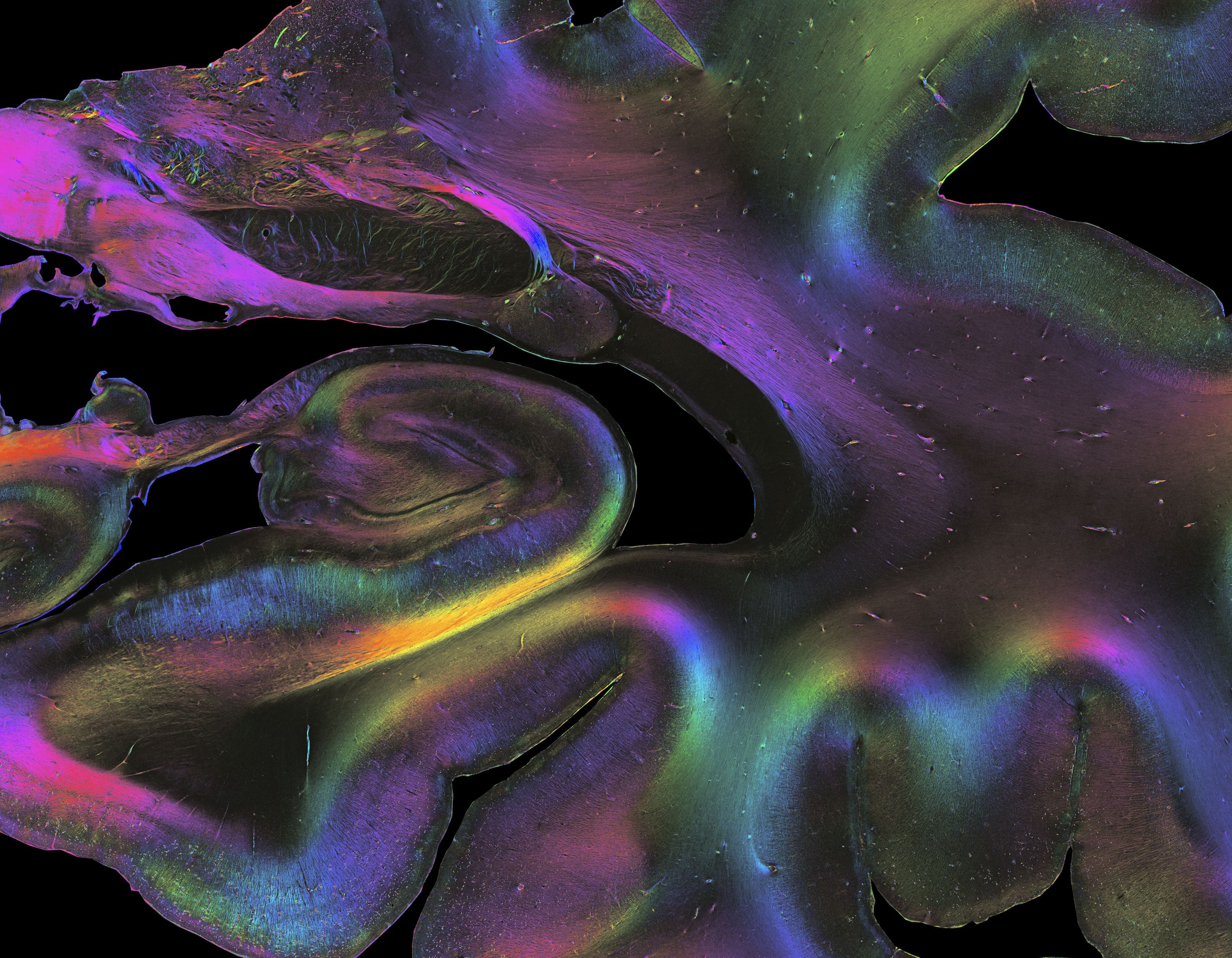
Detail of a human brain section showing the architecture of fibres down to single axons in the hippocampus, revealed by 3D Polarized Light Imaging. The hippocampus is critical for the creation, storage and recall of memories, and for connecting sensations and emotions to memories.
FLOWS
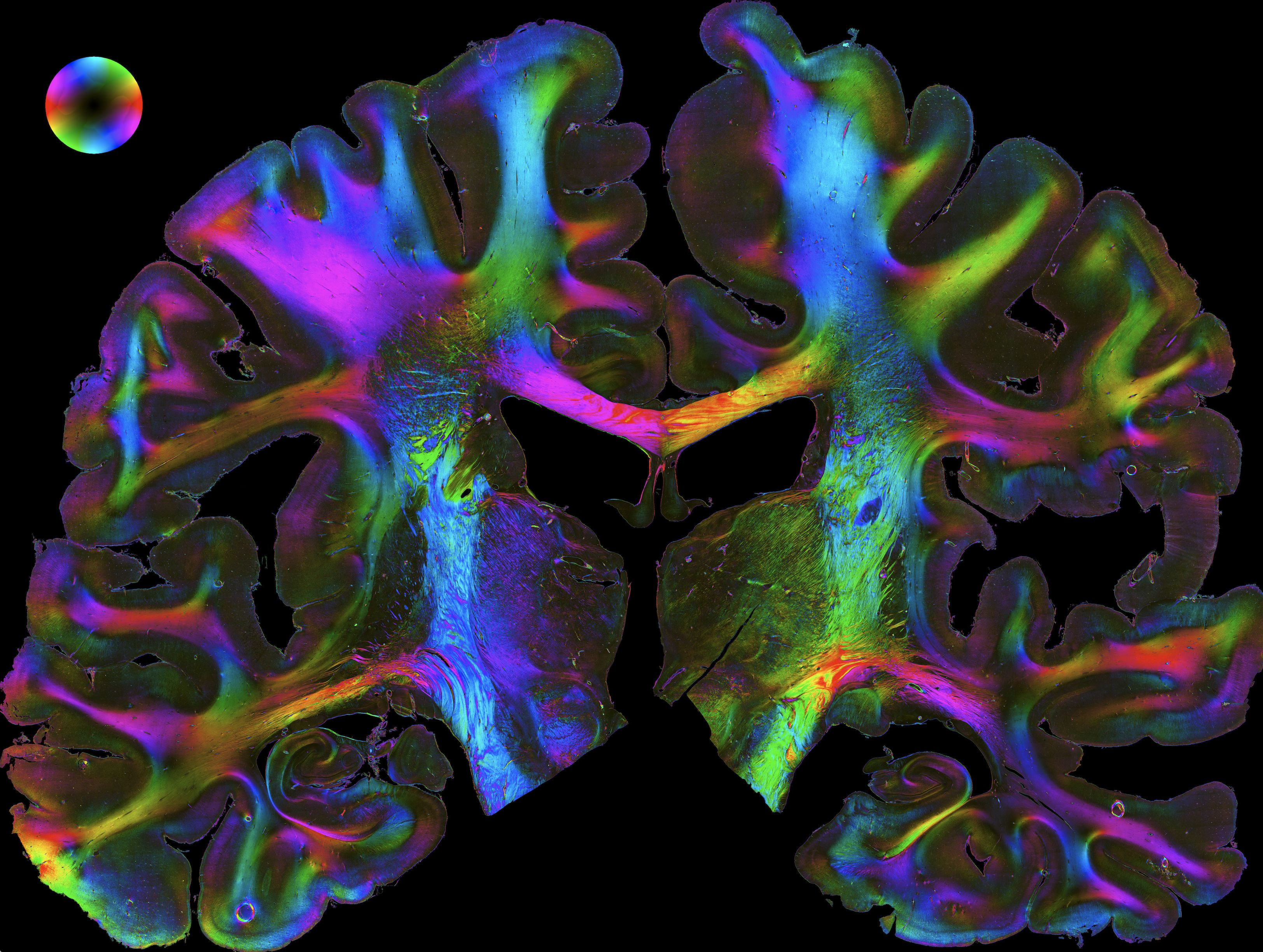
Fiber architecture of a coronal whole human brain section revealed with 3D Polarized Light Imaging. This imaging technique has been developed by the researchers to study the fiber orientation in a three-dimensional space.
Illuminating the brain
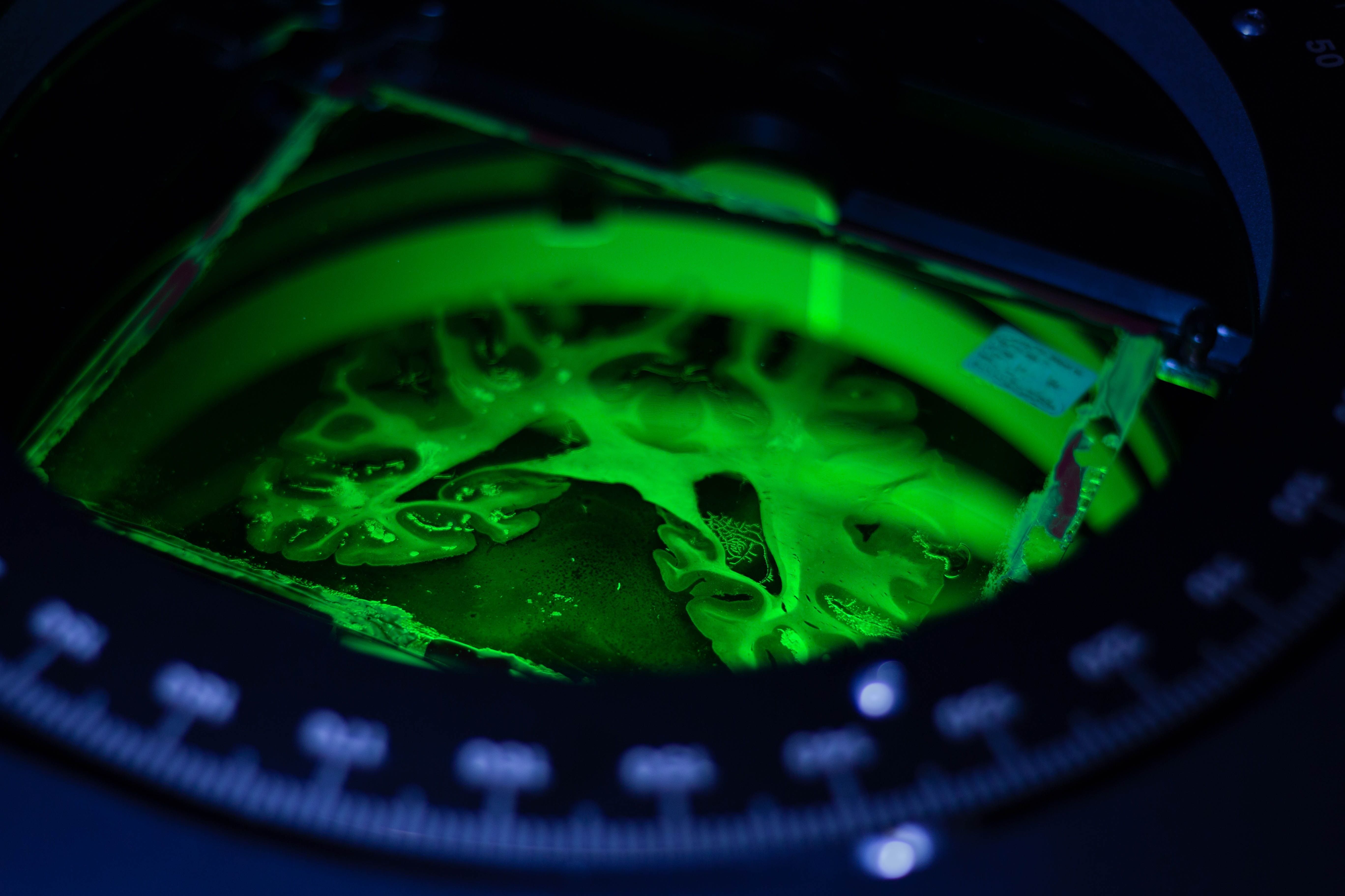
Brain section being imaged with polarized light to visualize fiber tracts at Forschungszentrum Jülich, in Germany. Each brain from individual donors is divided into many sections that are only a few micrometers thick.
PERSONAL BRAIN MODEL
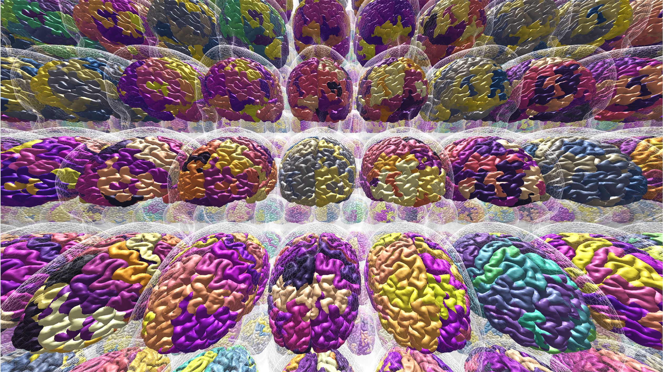
Computational models of the brain can be personalised for individual patients.
BRAIN TWINS
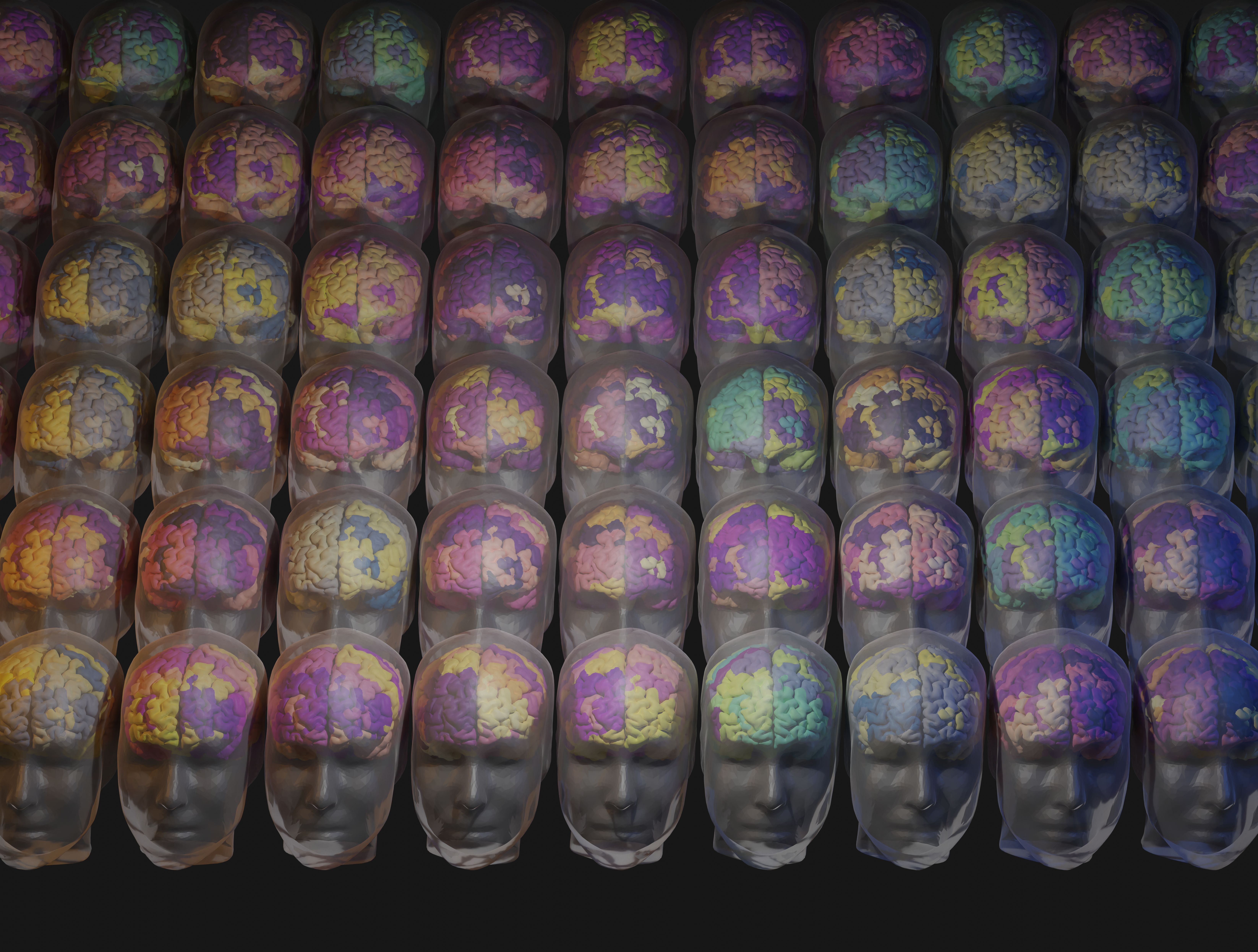
Virtualized brains displaying different brain states.
NETS

The Virtual Brain is a computational model exploiting the network nature of the brain. Each brain area is a network node, connected by links derived from the individual subject's own brain imaging data. Each person has their own virtual brain.
Check out the Interactive Atlas Viewer by The Virtual Brain!
REDWOODS
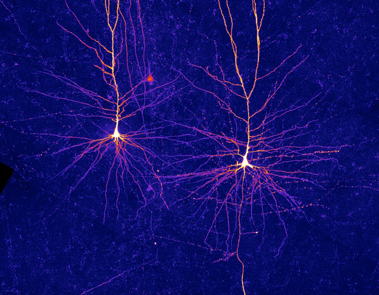
Neurons in layer 2/3 of the human neocortex, showing the tree-like branches called dendrites.
DEEP SPACE NEBULA
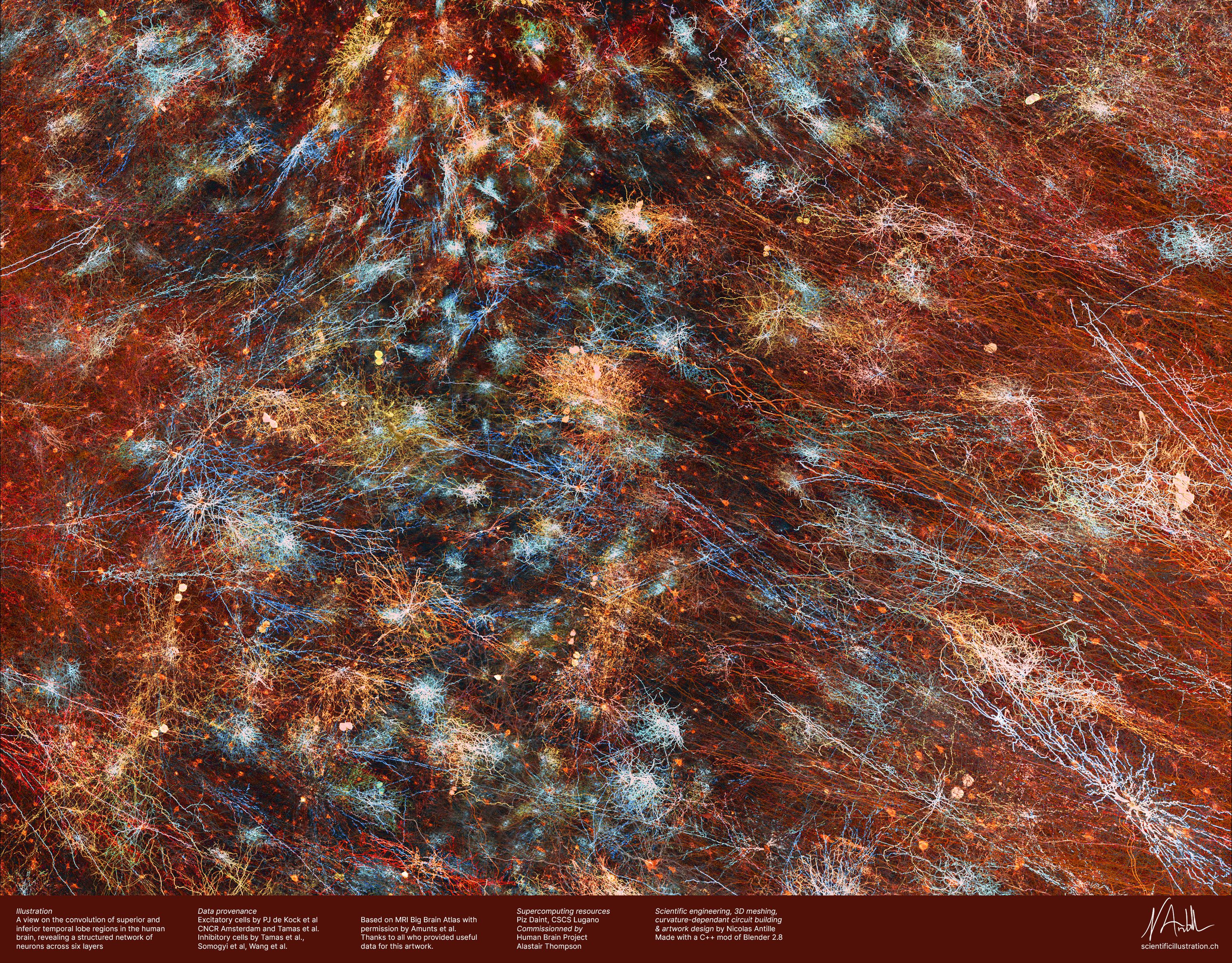
This scientific illustration gives a view of the temporal lobe regions in the brain.
HANDSHAKE
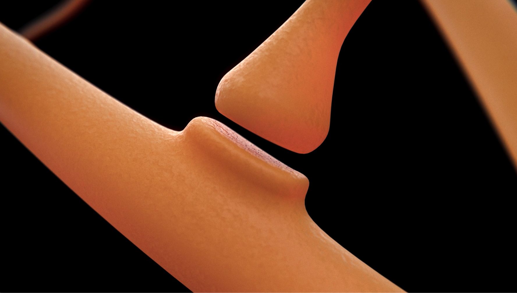
A 3D representation of a synapse, showing the interstitial space where the two neural cells exchange neurotransmitters.